See EEG in a whole new way.
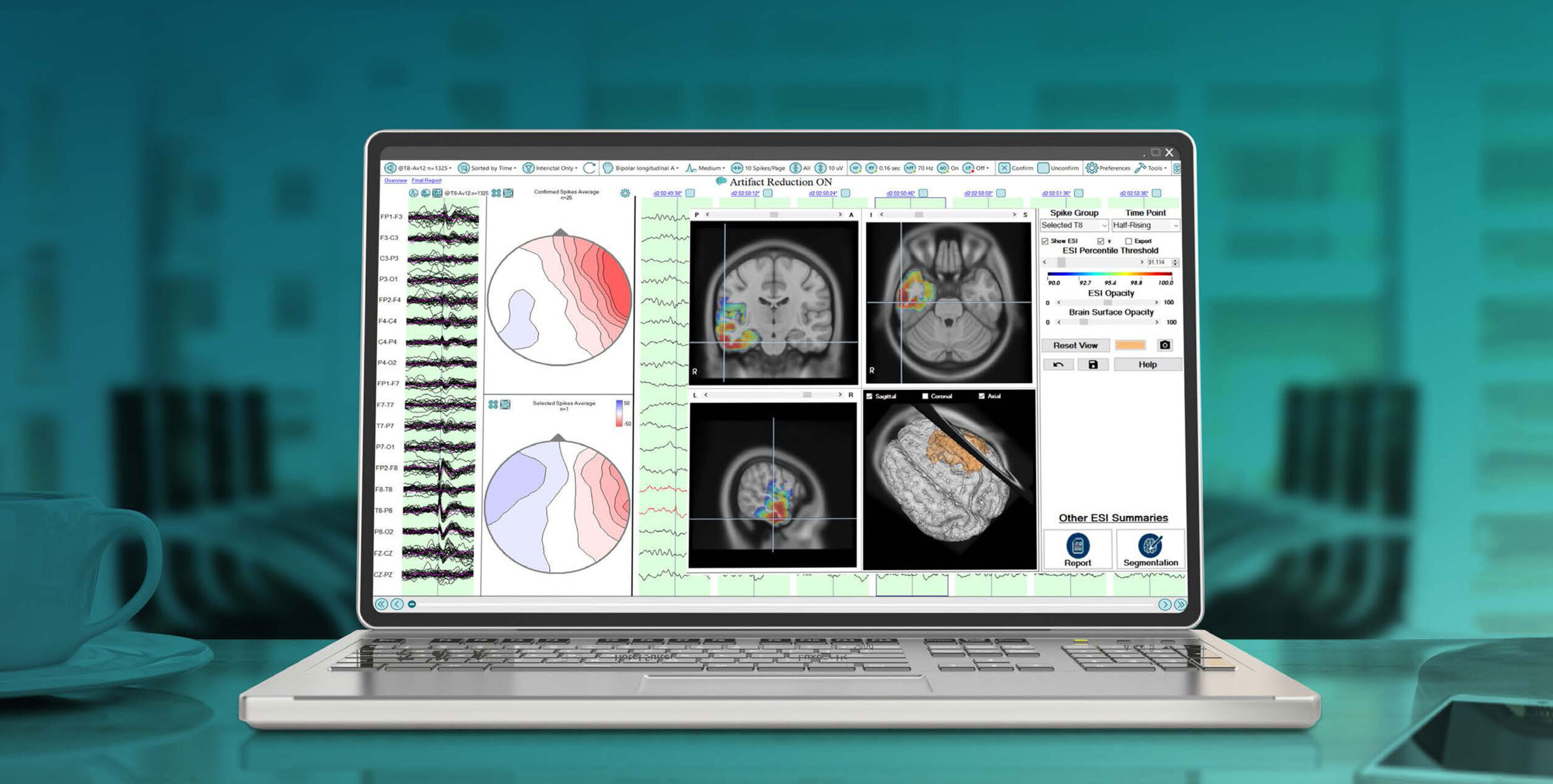
EEG Electrical Source Imaging (ESI) is a neuroimaging modality that uses a patient’s EEG data co-registered with their MRI to localize the source of the scalp EEG signals within the patient’s brain.
EEG Electrical Source Imaging has been shown to be both Accurate and Useful.
Now with Persyst, it’s Practical.
It’s Accurate.
Recent research shows that with the standard IFCN 25 EEG electrode array, ESI identifies epileptogenic brain regions well within the same range of accuracy as other conventional neuroimaging modalities such as MRI.1,2,3,4,5
One study, using a standardized methodology and clinician input, demonstrated that ESI accurately localized the epileptogenic zone in 78% of 41 consecutive surgical cases.1
Watch to learn about the history of EEG Electrical Source Imaging, our collaboration with Epilog and the methodological approach to Persyst ESI.
It’s Useful.
ESI significantly contributes to clinical decision-making, providing non-redundant information and is considered to be an important part of the presurgical evaluation for all patients considering epilepsy surgery. 6,7,8,9,12,13
Now with Persyst, it’s Practical.
Perform ESI using the software, equipment and skills you already have. 10,13
1 Baroumand AG, van Mierlo P, Strobbe G, Pinborg LH, Fabricius M, Rubboli G, et al. Automated EEG source imaging: A retrospective, blinded clinical validation study. Clin Neurophysi. (2018) 129:2403-2410.
2 Sharma P, Scherg M, Pinborg LH, Fabricius M, Rubboli G, Pedersen B, et al. Ictal and interictal electric source imaging in pre-surgical evaluation: a prospective study. European Journal of Neurology. (2018). 25(9):1154-1160.
3 Brodbeck V, Spinelli L, Lascano AM, Wissmeier M, Vargas MI, Vulliemoz S, et al. Electroencephalographic source imaging: a prospective study of 152 operated epileptic patients. Brain. (2011). 134:2887-2897.
4 Coito A, Biethahn S, Tepperberg J, Carboni M, Roelcke U, Seeck M, et al. Interictal epileptogenic zone localization in patients with focal epilepsy using electric source imaging and directed functional connectivity from low-density EEG. Epilepsia. (2019). 4(2):281-292.
5 Tatum WO, Rubboli G, Kaplan PW, Mirsatari SM, Radhakrishnan K, Gloss D, et al. Clinical utility of EEG in diagnosing and monitoring epilepsy in adults. Clinical Neurophysiology. (2018) 129:1056-1082.
6 Foged MT, Martens T, Pinborg LH, Hamrouni N, Litman M, Rubboli G, et al. Diagnostic added value of electrical source imaging in presurgical evaluation of patients with epilepsy: A prospective study. Clin Neurophys. (2020) 131:324-329.(2018) 129:1056-1082.
7 Megevand P, Vulliémoz S. Electric and Magnetic Source Imaging of Epileptic Activity. Epileptologie. (2013). 30:122-130
8 Rikir E, Koessler L, Gavaret M, Bartolomei F, Colnat-Coulbois S, Vignal JP, et al. Electrical source imaging in cortical malformation-related epilepsy: A prospective EEG-SEEG concordance study. Epilepsia (2014) 55(6):918-932
9 Beniczky S, Trinka E. Editorial: Source Imaging in Drug Resistant Epilepsy – Current Evidence and Practice. Frontiers in Neurology. (2020). 11(56).
10 Miron G, Baag T, Götz K, Holtkamp M, Vorderwülbecke BJ. Integration of interictal EEG source localization in presurgical epilepsy evaluation – A single-center prospective study. Epilepsia Open (2023) 8:877-887. 2023
11 Santalucia R, Carapancea E, Vespa S, Morrison EG, Baroumand AG, et al. Clinical added value of interictal automated electrical source imaging in the presurgical evaluation of MRI-negative epilepsy: A real-life experience in 29 consecutive patients. Epilepsy & Behavior (2023) 143:109229
12 Spinelli L, Baroumand AG, Vulliemoz S, Momjian S, Strobbe G, van Mierlo P, Seeck M. Semiautomatic interictal electric source localization based on long-term electroencephalographic monitoring: A prospective study. Epilepsia (2023) 64(4):951-961
13 Iachim E, Vespa S, Baroumand AG, Danthine V, Vrielynck P, et al. Automated electrical source imaging with scalp EEG to define the insular irritative zone: Comparison with simultaneous intracranial EEG. Clinical Neurophysiology (2021) 132(12):2965-2978
Incorporating ESI routinely for all evaluations has never been so accessible.
With Persyst, you can perform ESI using the resources and expertise you already have, making it practical and affordable.
- Integrated with all major EEG systems
- Works with standard 10-20 EEG arrays
- Scales up to 256 EEG channels
- Does not require additional hardware or special technical expertise
- Results in 2 business days
No ESI Solution is more accurate.
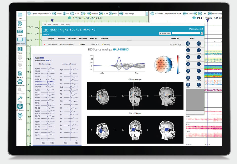
- Spike clusters from Persyst 15 spike detections and physician input
- Sources calculated at the onset, half-rising and peak of each spike cluster
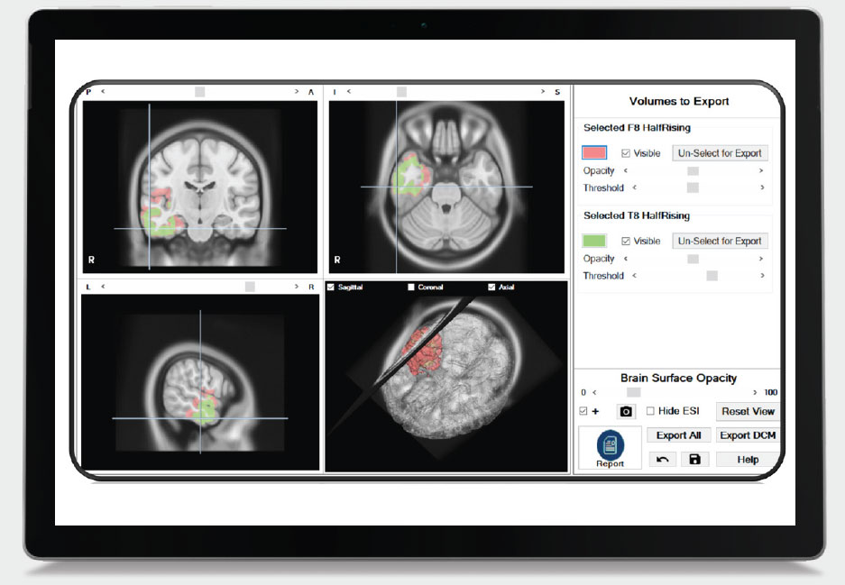
- Dynamically change the threshold of the ESI results
- Overlay and compare ESI results across clusters and time points
- Export ESI volumes to DICOM
- Current Density Reconstruction (sLORETA) combined with patient specific, high resolution FDM head models (Finite Difference Model) built from the patient’s own MRI, provides sub-lobar localization accuracy 10,11
Overview Page
The first page gives an overview of the spike clusters that have been detected, the lateralization of these clusters and at what time during the recording the spikes were detected.
Each spike cluster is color coded. This color is used throughout the report to denote this cluster.
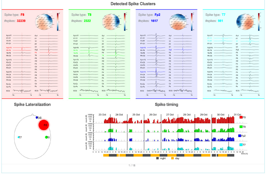
Spike detections / cluster
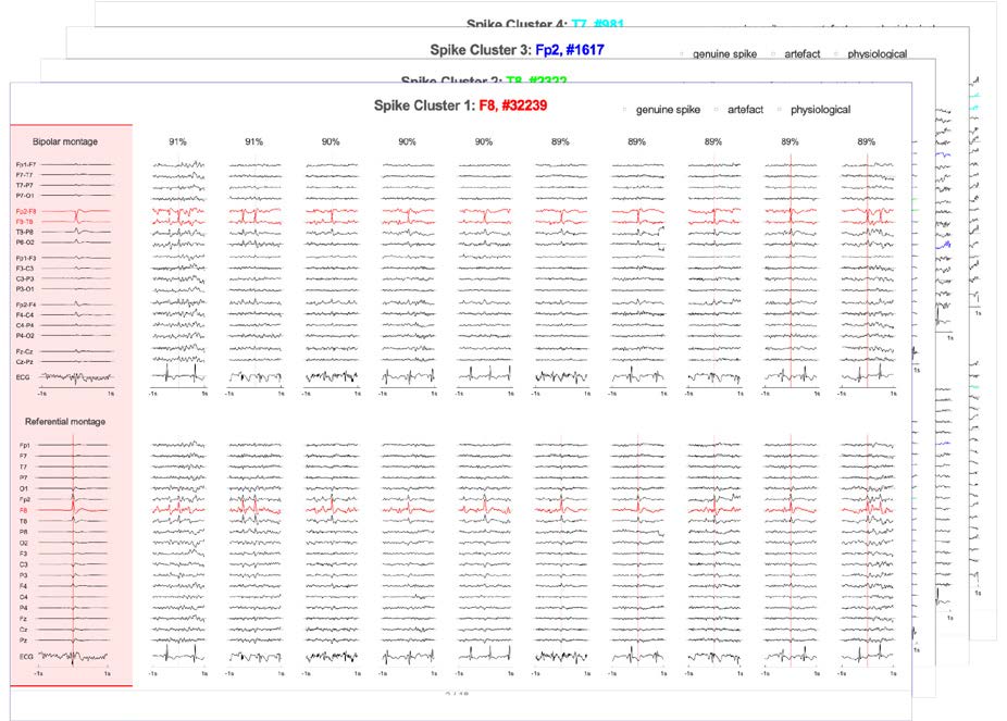
The next pages are grouped per cluster. The first page provides more detail about the detected spikes. Several individual spikes are displayed using bipolar and referential montages.
Spike localizations / cluster
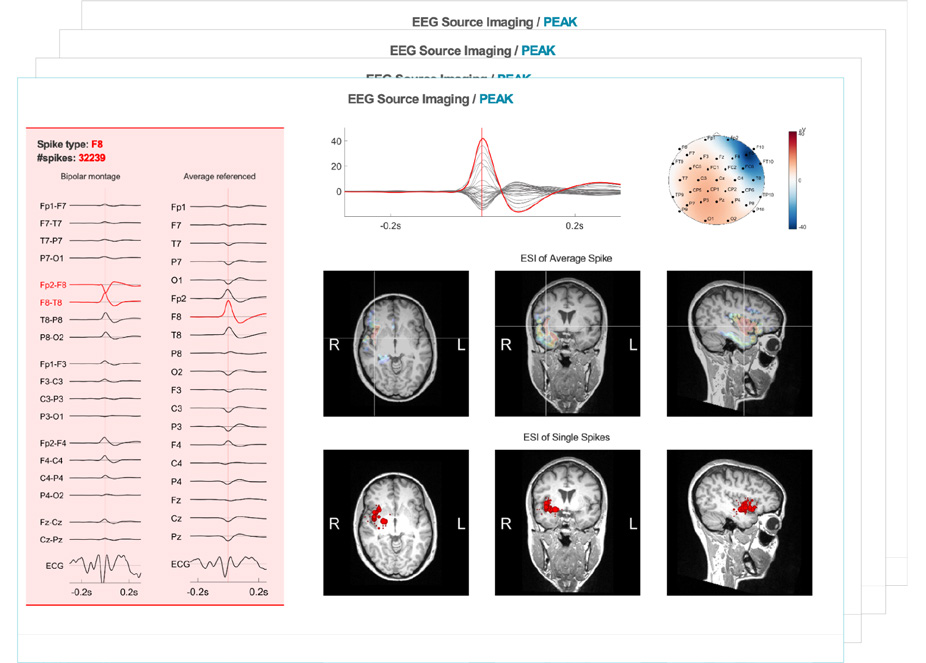
The following pages provide more detail about the source localized activity.
Source imaging results at the onset, half-rising, and peak are shown for the average spike waveform (top) in addition to the point maxima of the source imaging results for the individual spikes (below).
Head model
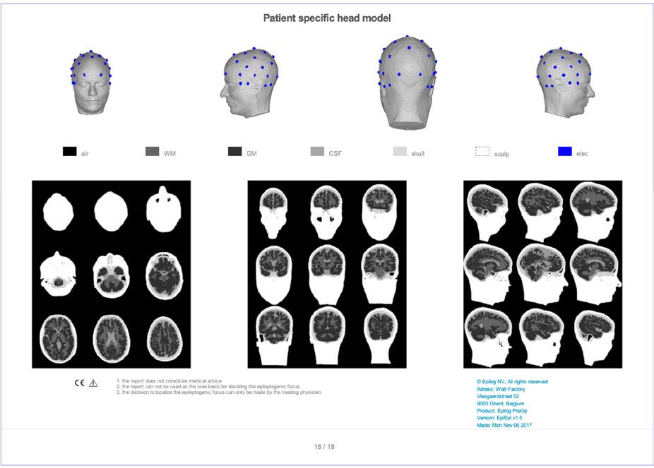
The last page provides information on the patient-specific head model that was used to perform source localization.


