
At the intersection of neuroscience and computer science,
Persyst is the worldwide leader in EEG software.
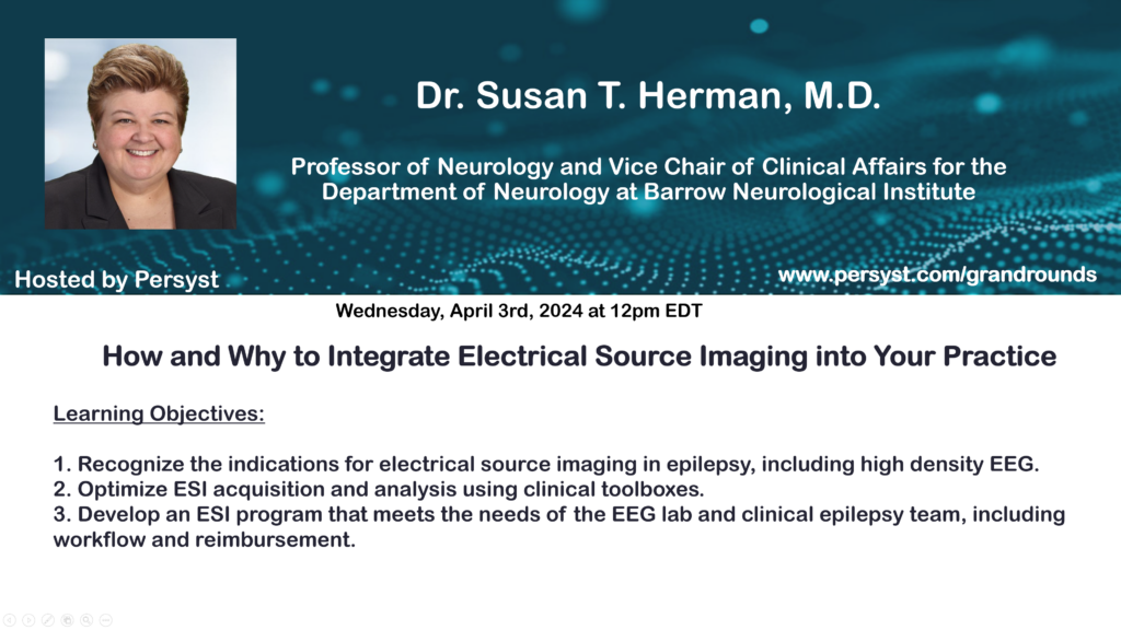
A series of online lectures
Lectures covering topics related to EEG and quantitative EEG.
")
The Second Edition of the Atlas of EEG in Critical Care by Dr. Lawrence Hirsch, Dr. Richard Brenner and Dr. Michael Fong is available.

Persyst participates in many conferences and events.
UT Houston Epilepsy Comprehensive Update & Board Review
Texas Medical Center
Houston, TX
May 8-9, 2024
Persyst | CARE
(Computer Assisted Review of EEG)
Persyst 14 EEG Review and Analysis Software provides the complete set of tools needed for C.A.R.E (Computer Assisted Review of EEG), resulting in accurate, efficient and rapid review of EEG data. Whether for high volume institutions or for community clinics, Persyst enables the highest level of comprehensive patient care when it comes to EEG monitoring and analysis.


Integrated with the EEG System you already use
Persyst is the only EEG trending and detection software that is integrated,
sold and supported by every major EEG manufacturer including
Micromed, Natus, Nihon Kohden, Cadwell, Compumedics and many more.


Persyst is the Standard of Care
* https://www.naec-epilepsy.org.
* As ranked by US News: https://health.usnews.com/best-hospitals/rankings/neurology-and-neurosurgery

Publications
Over the decades, Persyst has led the field of quantitative EEG for neurodiagnostics, delivering powerful tools into the hands of clinicians. Our success is in part due to a core commitment to research, the scientific validation of our methods in collaboration with our clinical partners, and the publication of our findings in peer-reviewed journals.
Most recently, our work validating the new Persyst 14 Seizure Detector was published by the Journal of Clinical Neurophsyiology. Findings include, “The Persyst 14 algorithm was statistically noninferior to the humans. For the first time, a seizure detection algorithm and human experts performed similarly.”